Implant placement in reduced bone volume
 For thirty years, implant placing has been a predictable and repetitive technique realized on young adults as well as elderly both in the maxilla and mandible with a success rate of more than 95% at 15 years old (Zarb and Schmitt 1990).
Vertical and transversal bone insufficiency is a common limitation of implant rehabilitation. It takes place to the detriment of the external cortical bone and lead to bone resorption and remodelling following extraction, infection, trauma or physiological. In this case, placing an implant is rather locally contraindicated and necessitates a prior treatment by means of onlay or vertical autogenous graft using either biomaterials or according to a guided bone regeneration technique with membrane or mesh. It requires a prolonged healing delay, from three to six months according to the type of the clinical case and more important post-operatory follow-ups.
We herein present the placement of M2I-MAGITECH implants as bone expanders of narrow ridges less or equal to 3mm in quality I or II of corticalized bone at the mandible and without any prior grafting at the maxilla. A second intervention is unnecessary enabling a global treatment cost reduction.
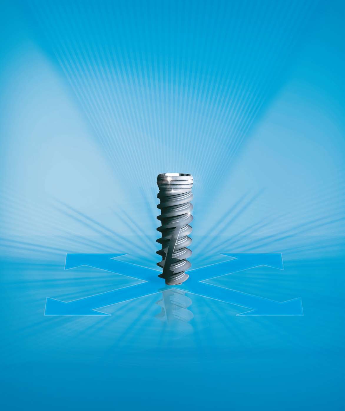
M2I-MAGITECH implants offer an ideal shape for bone expansion, very conical (better primary stability), with a reduced apical diameter from 1.5mm to 2.5mm according to the coronal diameter.
They are easily placed using a handle with insertion forces higher than 70N/cm. Their conicity enables lesser apical osseous constraints and a reduced bone heating by decreasing the number of drilling. This implant is of titanium alloy (TI6AL4 GRADE V) which mechanical features are 4 times higher (895 MPA traction resistance) than GRADE 1 COMMERCIALLY PURE TITANIUM (240 MPA) (Carr et al. 1997).
PRE-OPERATIVE ASSESSMENT
The pre-operative assessment which is a prerequisite fundamental step to any implantation act, commits the practitioner to study the relevance of the prosthetic project.
Any oral surgical act, simple or complex (bone surgery, implantology) requires a medical approach, based on a pre-operative assessment including notably a questioning and an exo- and endo-oral clinical exam. The ensuing observations and interrogations should lead us to reasonably prescribe complementary medical, biological or x-ray exams. Biological exams and the different expert advices will help evaluate the general condition of the patient, point out the contra-indications or the precautions to take when performing this act.
Medical imaging is a major important component in implantology where it evaluates the local and loco-regional condition. 2D x-ray evaluates infectious foyers, teeth that need extraction and implant feasibility.
Denta Scan and cone beam, 3D are used to assess bone deficiency. It confirms bone atrophy and specifies the available bone amount and the vestibular and palatine anatomical structures.
A moulding study with a resin prosthetic preparation helps visualize the final implant rehabilitation (number of teeth, occlusal ratios, implant positioning).
A radiographic guide is performed and will be modified into a surgical guide to allow positioning the implants according to three planes in space and to the final prosthesis desired by the patient.
SURGICAL TECHNIQUE
Interventions are performed under local anaesthesia after a peri and intra oral disinfection using 2% chlorhexidine or yellow betadine periorally or green betadine intraorally. The surgical zone is infiltrated with an adrenaline cartridge (DENTSPLY) thanks to its para-apical needle.
A full thickness crestal incision is made using a 15c scalpel blade extending the length of the edentulous area. It is continued with a slightly displaced buccal and lingual incision at the mandible and only buccaly at the maxilla.
The periosteal separation procedure is performed using molt periosteal elevator or a chompret syndesmotome. Using oschenbein scissors allow for scar mucous tissue dilacerations.
Osseous zone is then exposed delimitating the implant site. Mandibular bone crest presents a bucco-lingual thickness of 2.5mm represented thanks to a periodontal probe.
No further bone expansion or associated graft has been performed. A transverse osteotomy with crestal separation using a diamond disk or a piezosurgery according to a technique described by Vercellotti in 2001 has not been performed. Conical bone expander (Halberstam 2007) hasn’t been necessary to prepare the implant site.
At the mandible, two M2I-MAGITECH implants are to be placed in positions 34 and 44 (Clinical case 2).
Implant protocol MAGITECH M2I
A 0.5mm KOMET round bur is used to make a pre-hole according to the desired axis
A pilot drill of 1.5mm diameter is placed along the length of the predefined implant and then a MAGITECH 2mm diameter drill is used to finish the implant osteotomy to place the 3.3mm diameter M2I-MAGITECH implant.
Minimal drilling is sufficient even in highly corticalized bone as the apex diameter is only 1.5mm for a 3.3mm implant.
The implant and its titanium holder are transferred from their sterile capsule to be placed into the implant site using a ratchet wrench or a handle.
During the insertion into the implant site that was only prepared using two drills (1.5mm and 2mm), clinical case 2, M2I-MAGITECH implant penetrates the implant site (Figure 1 and 2) thanks to the patented angulated and double-conical threads. This implant enables a condensation but more importantly bone expansion without any risk of fracturing the external cortical bone.
M2I-MAGITECH implant may be inserted with insertion forces higher than 70N/cm without any apical osseous constraints because its apex is highly conical and allow for less bone heating as the apical osteotomy is reduced by 30%.
During its insertion, (clinical case, figure 3 and 4), we could observe a voussure and a cortical expansion with horizontal bone gain of 1.5mm to 2mm, required to securely placing a 3.3mm implant. This expansion is stable for a long term period even after uncovering the implant.
If the cortical bone surrounding the implant is less than 1.5mm or in presence of a crack, bone rearrangement maybe necessary using onlay synthetic biomaterials or xenografts (Bio-Oss Geistchlich) or autogenous bone grafts covered with Bio-Gide collagen membrane (Geischtlich) such as Janson (TLBM) or platelet derivatives (PRF) after preliminary blood test.
Bone healing delay following M2I-MAGITECH implant placement is only 6 weeks without filling as it presents a very high primary stability at insertion and motionlessness without micro-movements facilitating osteoblastic adhesion at the bone/implant interface.
In the present clinical cases, a bone rearrangement has been performed and all implants have been inserted and covered with a cover screw.
The second intervention was performed 12 to 14 weeks later because of bone rearrangement and after performing an x-ray.
Check-up 2D x-rays have been performed on the day of the intervention, 15 days, 1 month and 3 months later. A cone beam was realized 3 months later.
Healing screws and MAGITECH abutments were placed using a MAGITECH dynamometric wrench at 35N/cm to make the prosthesis.
At the maxilla presented in the CLINICAL CASE 1, the patient presented a series of interventions:
- Iliac onlay bone grafting, resorbed
- GBR technique, 3 months LATER
- FRIALITE IMPLANT (FRIADENT) in 11, overhanging at the buccal mucous, upper position, non utilizable aesthetically.
Crestal displaced and intra-sulcular incisions in palatine around 21 and 12 followed by a two vertical vestibular releasing incisions have allowed separating the muco-periosteal flap and giving access to the surgical site.
After excising the Friadent implant, the crest had a buccal-lingual bone thickness of 1.5mm which is not suitable to place M2I-MAGITECH implant.
Bone expansion by means of ridge splitting was developed by Tatum H in 1979 and resumed by many others (Scipioni 1994, Bravi 2007). This technique is used to sufficiently widen the ridge transversally. Two vertical corticotomies were outlined using a diamond saw (komet°) before placing the M2I-MAGITECH implant in order to perform the vestibular cortical bone expansion.
The M2I-MAGITECH implant’s double conicity allows succeeding an external cortical splitting safely, without fracturing the bone plate.
The simplicity of this technique is an advantage compared to autogenous bone grafting which presents variable results (A basa et al. 2004) necessitating a second painful operating site or biomaterial grafting according to the GBR technique (Dahlin 1988, Buser 1994) which is much more restricting to the patient.
M2I implant presents a high primary stability and an improved delay of bone healing. The second intervention is already possible after 6 weeks and prevents a prerequisite surgical bone rearrangement which reduces the global cost of the treatment.
POSTOPERATORY FOLLOW-UP
Antibiotherapy, Augmentin 1g in the morning and at night for 8 days, paracetamol-dextopropoxylene and chlohexidine are also given for a week.
After the placement of MAGITECH implants, a removable prosthesis emptied at the operated site will prevent any pressure on the operated zone. At the mandible (clinical case 2), healing abutments are placed, the gum is sutured in a higher position in order to delay epithelial migration according to a technique described by Prichard in 1957. Check-up x-rays were performed 15 days, 1 month and 4 months after the intervention.
An M2I-MAGITECH 15 degrees angulated abutment (clinical case 2) was placed at 18 weeks followed by a definitive ceramo-metal crown placement at 20 weeks.
The convex ridge profile obtained without mucous rearrangement is only due to osseous support after bone expansion and overfilling surrounding the implant which transformed this thin biotype into a thick biotype.
Discussion
The advantage of placing small diameters M2I-MAGITECH implants in narrow ridges is to eliminate any constraints related to a delicate surgery or inaccessible surgery for some. Its simple placement and exceptional primary stability allow reducing the healing period necessary for osteo-integration. Many studies (Mazor 2004, Romeo 2006 and Sennerby 2008) have shown a success rate over 96% for 3.3mm implants.
We could have performed a preliminary bone expansion to increase the crestal buccal-lingual diameter and suitably place the implant. However, this latter would have been too buried with a less functional prosthetic height and less esthetical result.
Using conic MAGITECH implants allow secure horizontal placement as the implant’s apex is pointy and has the same diameter as the last used drill instead of the diameter of the coronal implant. Its insertion into the bone enables a transversal bone gain and gives along a more convex volume and profile to soft tissues, thus improving the aesthetics.
Two studies (Sethi et al. 2000 and Simion et al. 1992) relate transversal bone expansion techniques at the maxilla and mandible with an immediate one-stage implant placement using a non-resorbable membrane according to the guided tissue regeneration principles.
Other authors, such as Pikos in 1992, use biomaterials to fill in the space after separating the bone wall with a postponed implant placement.
Bravi et al. 2007 has shown comparable results after alveolar split expansion and immediate 3.75mm and plus implant placement.
According to Princ and Piral in 2008, the splitting could hardly regain the too marked oblique feature of bone walls. A minimum of 3mm crestal width is required and presence of sponge bone is necessary between the external and internal cortical. High bone density makes splitting more difficult.
The most predictable technique for us was presented recently by Khoury et al. in 2010 at the mandible and consisted of a controlled osteotomy. In this case, stabilization and expansion are ensured using a lateral screw osteosynthesis.
Moreover, expansion doesn’t make up for a crestal delay of more than 3mm. Onlay autogenous bone grafting are thus necessary to improve the esthetic which necessitates a second operating site with a morbidity risk for the donor site.
According to Princ and Piral in 2008, grafting success criteria are:
Ø Stabilizing the graft using screw osteosynthesis
Ø Absence of hiatus between the graft and the receiving bed
Ø Absence of prominent edges on the graft which may disrupt gum healing
Ø Flap repositioning under tension
Ø A follow-up during the first weeks is crucial to the only bone graft survival
The MAGITECH implant safely tackles implant treatment in narrow ridges (less than 4mm) and prevents much heavier techniques that could be restricting to the patient. The intervention is simpler and the healing delay is reduced to six weeks compared to 16 weeks in grafting and expansion techniques and 24 weeks if the implant is postponed.
The transversal expansion technique is delicate and necessitates a skilful knowledge of the operating act, when splitting the bone, placing simultaneously the implant using bone expanders, before placing the implant. M2I-MAGITECH implant placement prevents such constraints and the preliminary expansion procedure.
In ridges less than 2mm, an expansion technique by transversal osteotomy is required to increase the diameter to 1-2mm, without using an expander as the MAGITECH implant is able to play that role. Placing other implants would have required a prerequisite bone rearrangement, more restricting, with a longer healing delay and more important follow-up operations.
Conclusion
Placing M2I-MAGITECH implants in narrow ridges higher than 2mm limits the resort to bone sampling which necessitates a second operation site or the distraction technique which prevents eventual morbidity of the donor site.
Bone expansion techniques are not simple to realize and requires perfect knowledge of the surgical skill.
Using conic implants allow bone insertion with less osseous constraint and less heating thanks to its reduced apical diameter by 30% compared to cylindrical implants.